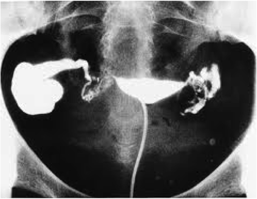
This test allows us to get a picture of the inner shape of the uterus and the patency of the Fallopian tubes.
Exam description:
A gynaecological examination is carried out on the radiology table, during which a speculum is inserted to be able to see the cervix of the uterus. A clamp is then placed around the neck of the uterus and an oily liquid inserted into the uterus using a special cannula, which then fills up the interior cavity of the uterus and runs out through the Fallopian tubes and reaches the woman’s abdominal cavity. Meanwhile, X-rays are taken.
Practical info : the timing of the test is extremely important. The test is carried out after the end of the period and before ovulation. This avoids exposing a fertilised egg to X-rays. You should therefore make an appointment for this test in consultation with your doctor.
Report to the gynaecology consultation 15 minutes before the test. An ultrasound is performed to rule out ovulation. The secretary will ask you to take a urine pregnancy test. You will then go to the radiology department. Your fertility doctor performs the gynaecological part and the radiologist takes the X-rays. Your doctor will generally prescribe you a preventive antibiotic to kill any bacteria present on the cervix of the uterus that could penetrate via the test.
The test sometimes causes slight discomfort, but in most cases it is not that bad. A painkiller before the test (e.g. brufen) can be useful, but is not required in most cases.